


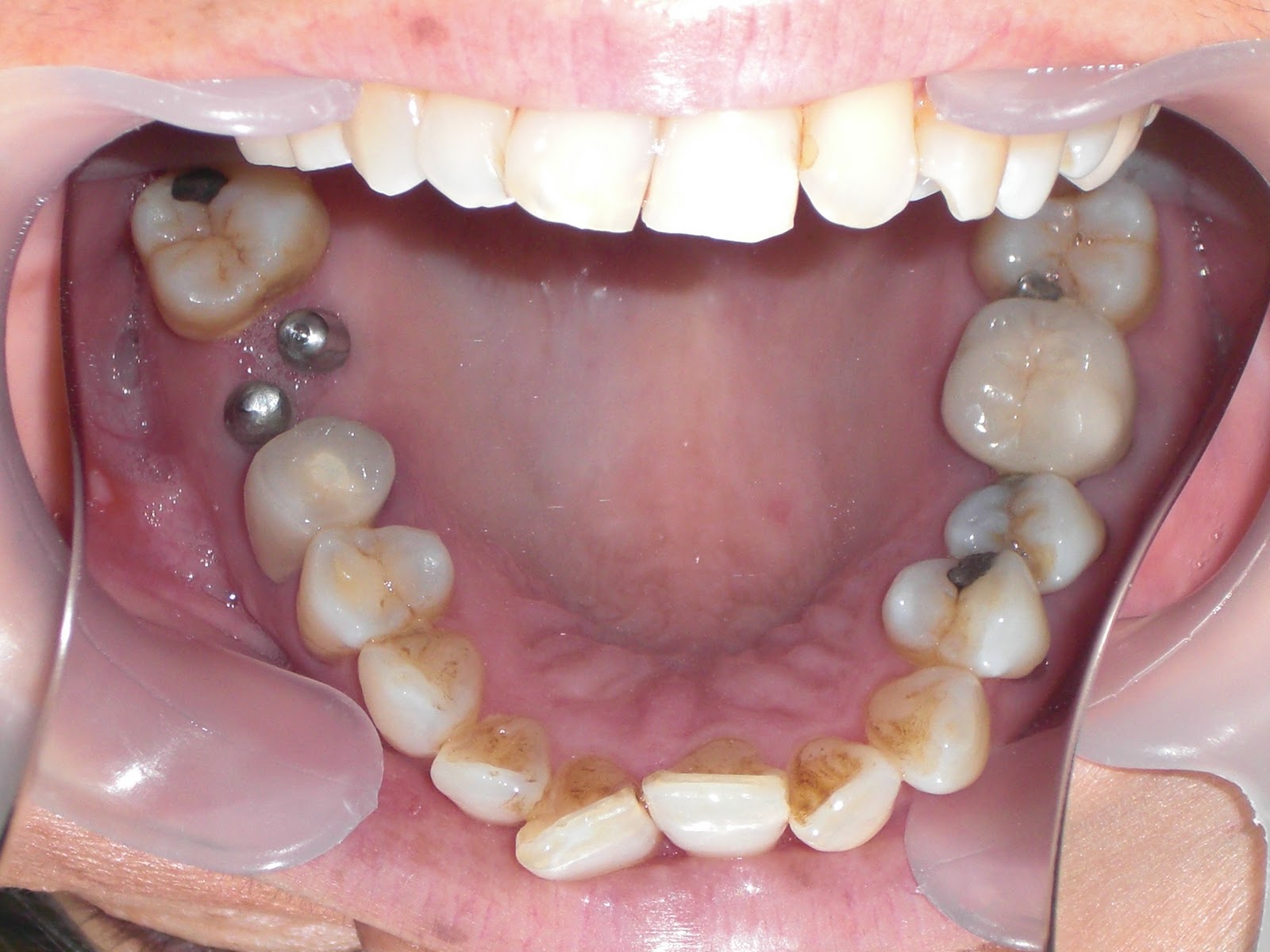

Notice the "CLEANING GROOVE" between the two abutment holes. This is to facilitate the threading of a bridge cleaner and floss in between and thus the undersurface of the crown can be flossed right to the surface of the implants. The ability to floss thus and the smallness of the emergence margin of the mini will arguably prevent any peri-implantitis in the long run.
In contrast, a conventional implant with an aesthetic emergence profile that includes a large emergence margin cannot be flossed all the way to the surface of the implant proper. The emergence profile emerges out of a volcanic-like crater in the gums. The surface of the crater is usually slightly inflamed and together with the large surface will arguably be more susceptible to peri-implantitis as compared to a mini dental implant. Examine the pics below.


Let's continue with the replacement of the upper molar by copying the anatomical/biologically ergonomical/natural/no-brainer/obviously correct positions of the roots.


The upper right first molar was deemed unsavable and extracted. After a short healing period, 2 minis were placed. One on the palatal wall of the sinus and the other on the buccal wall of the sinus. The divinely ordained and inarguably best anatomical positions of the palatal and disto-buccal roots were copied by the two mini dental implants. The undersurface of the crown was designed with a"CLEANING GROOVE" to enable threading of a dental floss and flossing.
Hi! How long do you usually wait after an extraction before placing a mini?
ReplyDeleteAlso after placement of a mini do you wait for some sort of integration before cementing a crown? :)